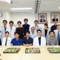
One of our medical student, Dr. Yuichiro Koga has published his first paper in the official journal of Japan Neurosurgical Society, Neurol Med Chiri (Tokyo) Case Report J today. He has passed National Examination for Medical Practitioners in this March and will start his residency program in this April. We are ready to fully support him to be an excellent neurosurgeon in very near future!
Boys and girls, be ambitious!
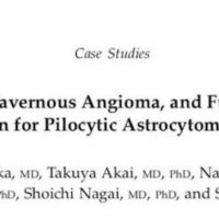
Yuichiro Koga, Saori Hamada, Hisayasu Saito, Takuya Akai, Satoshi Kuroda:
Intracranial, Intra-parenchymal Capillary Hemangioma – Case Report –
Neurol Med Chiri (Tokyo) Case Report J March 24, 2020 [Epub ahead of print]
ABSTRACT
We report a very rare case of intracranial capillary hemangioma. This 15-year-old girl complained of pulsating
headache in the temple area that aggravated with change
of body positions. This headache usually lasted for 5 min
and resolved without any treatment. Preoperative computed
tomography (CT) and magnetic resonance imaging
(MRI) strongly suggested cavernous hemangioma in the
right deep parietal lobe. She underwent complete resection
of the tumor through right parietal craniotomy. Postoperative
course was uneventful. Histologic examinations
demonstrated a densely grown numerous capillary-like
vascular structure with endothelial cells, hemosiderin
deposition, and hemorrhage. Intracranial, intra-parenchymal
capillary hemangioma is a very rare vascular tumor or
tumor like lesions. Only four cases with intracranial, intraparenchymal capillary hemangioma were reported previously. Differential diagnosis includes other vascular
tumors such as cavernous hemangioma, but it is not so
easy to differentiate capillary hemangioma from other
lesions. Therefore, surgical excision and histologic diagnosis
would be important to diagnose it if possible.