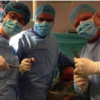
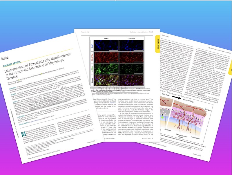
当科の大学院生・山本修輔先生の研究論文が米国脳卒中協会のStroke誌 (impact factor=7.914)に掲載されました。もやもや病に対する脳血行再建術を実施する際、脳表クモ膜の小片を採取して免疫染色を施したところ、もやもや病ではクモ膜の線維芽細胞(fibroblast)数が増加しているのみならず、これらの多くが筋線維芽細胞(myofibroblast)に分化していること、もやもや病のクモ膜ではコラーゲン線維が通常よりも多く形成されていることが初めて明らかとなりました。同時に、もやもや病の脳脊髄液ではその分化に関与しているサイトカインであるbFGF, HGF, TGF-bが高濃度に含まれていることも明らかとなりました。
近年、共同研究者の笹原正清先生、山本誠士先生らの研究によって、筋線維芽細胞(myofibroblast)は創傷の治癒や血管新生に大きな役割を果たしていることが判明しており、われわれはこれらの現象が、もやもや病で特異的に認められる側副血行路や間接バイパスの形成に深く関与していると考えています。
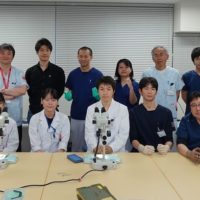
今後も、もやもや病の基礎研究・臨床研究を推進することで、次世代の研究者を育成して、もやもや病の患者さんに少しでも貢献したいと思います。今回も笹原正清先生、山本誠士先生には多大なご理解とご助力を賜わりました。この場をお借りして御礼申し上げます。
Boys and girls, be ambitious!
A Voyage to Depth of Neuroscience Vol. 85
A research article by Dr. Shusuke Yamamoto, a PhD student at our department, was published in Stroke (impact factor=7.914), a journal of the American Stroke Association. Immunostaining of a small piece of the arachnoid membrane of the brain surface during surgical revascularization for moyamoya disease revealed that not only the number of fibroblasts in the arachnoid membrane was increased in moyamoya disease, but many of them differentiated into myofibroblasts. Furthermore, collagen fibers are formed in higher amounts than usual in the arachnoid membranes of patients with moyamoya disease. At the same time, we also found that the cerebrospinal fluid of moyamoya disease contains high concentrations of bFGF, HGF, and TGF-b, which mediates the differentiation process.
Recently, myofibroblasts have been found to play a major role in wound healing and angiogenesis by our collaborators, Prof. Masakiyo Sasahara and Dr. Seiji Yamamoto, and we believe that these phenomena are deeply involved in the formation of collateral blood vessels and indirect bypasses, which are specifically observed in moyamoya disease.
By promoting basic and clinical research on moyamoya disease, I would like to continue to train the next generation of researchers and contribute to patients with moyamoya disease.
Boys and girls, be ambitious!
Differentiation of Fibroblasts Into Myofibroblastsin the Arachnoid Membrane of Moyamoya Disease
Shusuke Yamamoto, Seiji Yamamoto, Takuya Akai, Masakiyo Sasahara, Satoshi Kuroda
BACKGROUND: Moyamoya disease (MMD) is a very specific disorder in terms of spontaneous development of extracranialto-intracranial collateral circulation through the dura mater, but the underlying mechanisms are unclear. This study aimed toinvestigate the role of the arachnoid membrane in this unique angiogenesis in MMD.
METHODS: A piece of arachnoid membrane and 1- to 2-mL cerebrospinal fluid were simultaneously harvested duringsurgery from 26 patients with MMD. The specimens were also collected during surgery as the controls from 6 patientswith atherosclerotic carotid artery diseases. The arachnoid membrane was subjected to immunohistochemistry and thecerebrospinal fluid was used to measure the concentration of cytokines using ELISA.
RESULTS: The number of cells positive for PDGFR (platelet-derived growth factor receptor) α was significantly higher inMMD than in the controls (5.4±3.1 versus 2.3±2.1 cells/field; P=0.02). The results were same in PDGFRβ-positive cells(10.1±4.6 versus 4.8±2.8; P=0.01) and α-SMA (alpha-smooth muscle actin)–positive cells (8.8±3.1 versus 2.0±2.5;P<0.01). On multicolor immunofluorescence, 80.5±15.6% of cells positive for PDGFRα in MMD also expressed α-SMA,being significantly higher than 14.6±7.2% in the controls (P<0.01). The density of collagen in the arachnoid membrane wassignificantly higher in MMD than in the controls (60.3±15.0% versus 40.1±15.3%; P<0.01). In MMD, advanced diseasestage was significantly associated with a larger number of α-SMA–positive cells in the arachnoid membrane (P=0.04). OnELISA, the cerebrospinal fluid concentrations of bFGF (basic fibroblast growth factor), HGF (hepatocyte growth factor), andTGF (transforming growth factor)-β1 were significantly higher in MMD than in the controls.
CONCLUSIONS: Based on these findings, MMD may elevate the concentrations of angiogenic factors in the cerebrospinalfluid and then promote the proliferation of fibroblasts in the arachnoid membrane and their differentiation intomyofibroblasts, which may, in turn, enhance the production of collagen essential for spontaneous collateral formationacross the arachnoid membrane.