当科の研修医、城 泰輔先生の論文がWorld Neurosurgery誌に掲載されました。過去、もやもや病を対象とした横断的研究では、側頭骨の頚動脈管が縮小していると報告されています。しかし、これまで縦断的研究は実施されていませんでした。
今回、われわれは少なくとも5年間経過観察した成人もやもや病のうち、経過観察中に病期が進行した症例を対象に、経時的に頚動脈管径を測定したところ、病期進行の6ヶ月後から頚動脈管の径が縮小し始め、病期進行から5年後には初期値の85%まで縮小することを初めて明らかにしました。
これまで城先生は症例報告 2編(日本語 1編、英語 1編)を発表していますが、今回の論文は初めてのcase series reportです。診療にも研究にも、さらに貪欲に取り組んでほしいですね。
Boys and girls, be ambitious!
A Voyage to Depth of Neuroscience Vol. 82
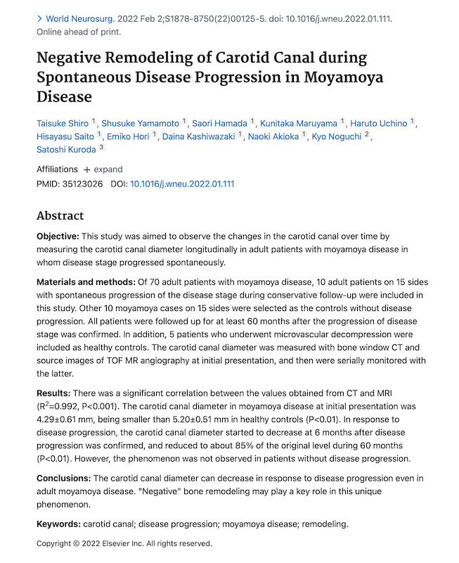
Negative Remodeling of Carotid Canal during Spontaneous Disease Progression in Moyamoya Disease
Taisuke Shiro, Shusuke Yamamoto, Saori Hamada, Kunitaka Maruyama, Haruto Uchino, Hisayasu Saito, Emiko Hori, Daina Kashiwazaki, Naoki Akioka, Kyo Noguchi, Satoshi Kuroda
Abstract
Objective: This study was aimed to observe the changes in the carotid canal over time by measuring the carotid canal diameter longitudinally in adult patients with moyamoya disease in whom disease stage progressed spontaneously.
Materials and methods: Of 70 adult patients with moyamoya disease, 10 adult patients on 15 sides with spontaneous progression of the disease stage during conservative follow-up were included in this study. Other 10 moyamoya cases on 15 sides were selected as the controls without disease progression. All patients were followed up for at least 60 months after the progression of disease stage was confirmed. In addition, 5 patients who underwent microvascular decompression were included as healthy controls. The carotid canal diameter was measured with bone window CT and source images of TOF MR angiography at initial presentation, and then were serially monitored with the latter.
Results: There was a significant correlation between the values obtained from CT and MRI (R2=0.992, P<0.001). The carotid canal diameter in moyamoya disease at initial presentation was 4.29±0.61 mm, being smaller than 5.20±0.51 mm in healthy controls (P<0.01). In response to disease progression, the carotid canal diameter started to decrease at 6 months after disease progression was confirmed, and reduced to about 85% of the original level during 60 months (P<0.01). However, the phenomenon was not observed in patients without disease progression.
Conclusions: The carotid canal diameter can decrease in response to disease progression even in adult moyamoya disease. “Negative” bone remodeling may play a key role in this unique phenomenon.