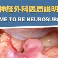
当科の柏崎大奈先生が書いた頚動脈狭窄症に関する論文が欧州脳神経外科学会誌「Acta Neurochirurgica (Wien)」誌に掲載されました。
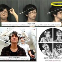
もやもや病では脳循環動態が不良な領域でMRI FLAIR imagingにて脳表の動脈が高信号として描出される”ivy sign”は、広く知られてるようになりました。
しかし、今回の画像解析によって、頚動脈狭窄症においても類似した所見が二つのメカニズムによって出現することが判明しました。
1)高度狭窄による血行力学的脳虚血(約40%)
2)不安定プラークによる微小塞栓(約60%)
われわれは、この所見をhigh-intensity vessel sign on FLAIR imaging (HVS)と命名しました。このHVSは、頚動脈狭窄症を有する症例において、脳梗塞の発症や再発を予知できる画像マーカーになると考えています。
https://pubmed.ncbi.nlm.nih.gov/32458404/…
A Voyage to Depth of Neuroscience Vol. 72
Dr. Daina Kashiwazaki, one of our staffs, has published his clinical research paper on carotid artery stenosis in Acta Neurochirurgica (Wien) on May 26, 2020.
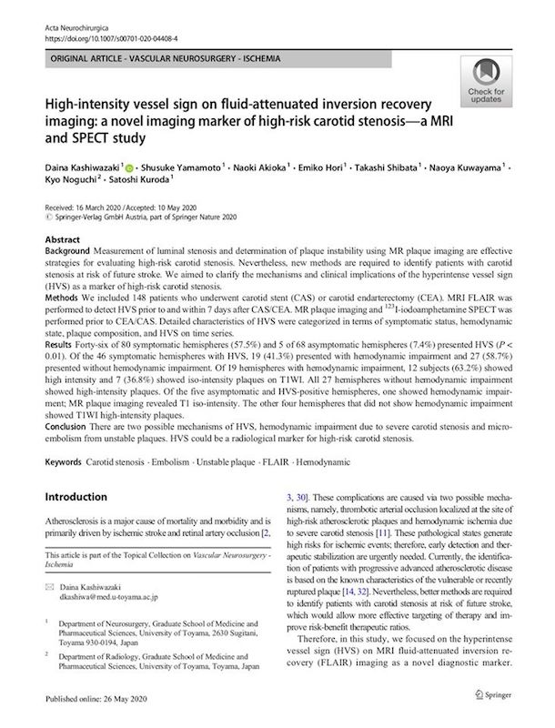
Kashiwazaki D, Yamamoto S, Akioka N, Hori E, Shibata T, Kuwayama N, Noguchi K, Kuroda S.
High-intensity Vessel Sign on Fluid-Attenuated Inversion Recovery Imaging: A Novel Imaging Marker of High-Risk Carotid Stenosis-A MRI and SPECT Study
Acta Neurochir (Wien). 2020 May 26. Online ahead of print.
Abstract
Background: Measurement of luminal stenosis and determination of plaque instability using MR plaque imaging are effective strategies for evaluating high-risk carotid stenosis. Nevertheless, new methods are required to identify patients with carotid stenosis at risk of future stroke. We aimed to clarify the mechanisms and clinical implications of the hyperintense vessel sign (HVS) as a marker of high-risk carotid stenosis.
Methods: We included 148 patients who underwent carotid stent (CAS) or carotid endarterectomy (CEA). MRI FLAIR was performed to detect HVS prior to and within 7 days after CAS/CEA. MR plaque imaging and 123I-iodoamphetamine SPECT was performed prior to CEA/CAS. Detailed characteristics of HVS were categorized in terms of symptomatic status, hemodynamic state, plaque composition, and HVS on time series.
Results: Forty-six of 80 symptomatic hemispheres (57.5%) and 5 of 68 asymptomatic hemispheres (7.4%) presented HVS (P < 0.01). Of the 46 symptomatic hemispheres with HVS, 19 (41.3%) presented with hemodynamic impairment and 27 (58.7%) presented without hemodynamic impairment. Of 19 hemispheres with hemodynamic impairment, 12 subjects (63.2%) showed high intensity and 7 (36.8%) showed iso-intensity plaques on T1WI. All 27 hemispheres without hemodynamic impairment showed high-intensity plaques. Of the five asymptomatic and HVS-positive hemispheres, one showed hemodynamic impairment; MR plaque imaging revealed T1 iso-intensity. The other four hemispheres that did not show hemodynamic impairment showed T1WI high-intensity plaques.
Conclusion: There are two possible mechanisms of HVS, hemodynamic impairment due to severe carotid stenosis and micro-embolism from unstable plaques. HVS could be a radiological marker for high-risk carotid stenosis.