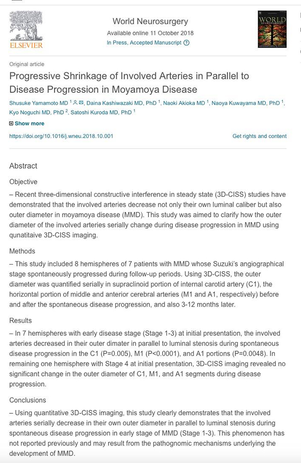
当科のレジデント、山本修輔先生の論文「もやもや病の病期進行に合わせて罹患動脈の外径も縮小する」がWorld Neurosurgery誌に掲載されました。
近年の研究によって、もやもや病の罹患動脈では内径のみならず外径も縮小することは広く知られつつありますが、病期の進行によって外径がどのように変化するのかについての研究はこれまでなされてきませんでした。もやもや病の病態解明、診断精度の向上に役立つ研究になれば、と考えています。
A Voyage to Depth of Neuroscience Vol. 54
One of our residents, Dr. Shusuke Yamamoto published a paper on serial changes in outer diameter during spontaneous disease progression in moyamoya disease.
Yamamoto S, Kashiwazaki D, Akioka N, Kuwayama N, Noguchi K, Kuroda S.
World Neurosurg. 2018 Oct 10. [Epub ahead of print]
Progressive Shrinkage of Involved Arteries in Parallel to Disease Progression in Moyamoya Disease.
Abstract
OBJECTIVE:
– Recent three-dimensional constructive interference in steady state (3D-CISS) studies have demonstrated that the involved arteries decrease not only their own luminal caliber but also outer diameter in moyamoya disease (MMD). This study was aimed to clarify how the outer diameter of the involved arteries serially change during disease progression in MMD using qunatitaive 3D-CISS imaging.
METHODS:
– This study included 8 hemispheres of 7 patients with MMD whose Suzuki’s angiographical stage spontaneously progressed during follow-up periods. Using 3D-CISS, the outer diameter was quantified serially in supraclinoid portion of internal carotid artery (C1), the horizontal portion of middle and anterior cerebral arteries (M1 and A1, respectively) before and after the spontaneous disease progression, and also 3-12 months later.
RESULTS:
– In 7 hemispheres with early disease stage (Stage 1-3) at initial presentation, the involved arteries decreased in their outer dimater in parallel to luminal stenosis during spontaneous disease progression in the C1 (P=0.005), M1 (P<0.0001), and A1 portions (P=0.0048). In remaining one hemisphere with Stage 4 at initial presentation, 3D-CISS imaging revealed no significant change in the outer diameter of C1, M1, and A1 segments during disease progression.
CONCLUSIONS:
– Using quantitative 3D-CISS imaging, this study clearly demonstrates that the involved arteries serially decrease in their own outer diameter in parallel to luminal stenosis during spontaneous disease progression in early stage of MMD (Stage 1-3). This phenomenon has not reported previously and may result from the pathognomic mechanisms underlying the development of MMD.